
Maybe you noticed a discolored patch in the hospital, or a strange spot appeared a few weeks later. It is easy to worry, but birthmarks and skin variations are incredibly common. They are part of what makes your baby unique.
Some marks vanish on their own, while others stick around for life. In rare cases, a spot might signal an underlying condition.
Let’s break down everything you need to know about baby skin discoloration, from harmless patches to signs that need a doctor’s attention.
Key Takeaways
- Birthmarks come in two main categories: vascular (blood vessels) and pigmented (cell clusters).
- Common temporary issues include jaundice (yellowing) and salmon patches (“stork bites”).
- Watch for “six or more” Cafe au lait spots, as this can indicate a genetic condition.
- Most birthmarks are harmless, but rapid changes or bleeding require a pediatrician’s check.
Baby Skin Color 101
In our heads, we picture a newborn as a chubby-cheeked wonder with flawless skin. Reality is often a little messier.
Fresh from the womb, a newborn is often purple and slimy. They may also be covered in a white, cheese-like substance called vernix caseosa. This protects their skin in the amniotic fluid.
Because a baby’s skin is paper-thin, environmental changes play a huge role in their color.
You might notice their hands and feet look bluish. This is called acrocyanosis. It is normal in the first few days as their circulation figures itself out. If they are cold, you might see “mottling,” which looks like a lacy, marble pattern on their legs or chest.
Conversely, their cheeks will get red and flushed if they are hot or crying.
Permanent Skin Tone
Newborns sometimes have a lighter complexion than you expected. Pigment takes time to develop.
Some babies take up to six months or longer to settle into their permanent skin color. Genetics play the biggest role here, so looking at mom and dad is your best clue.
Pro Tip
Brown Spots and Discoloration
Brown spots can be more than just cute freckles. It is not uncommon to find a few when bathing your baby. Don’t worry; most baby spots are benign.
Here is what might be causing those darker patches:
Cafe Au Lait Macules
Cafe au lait macules (CALMs) get their name from the French term for “coffee with milk” because of their light brown color.
These flat spots can appear anywhere on a baby’s body. They are usually present from birth but can develop in the first few years.
Quick facts about CALMs:
- Found in about 3% of infants.
- Typically random, uneven shapes.
- Range from tiny dots to larger patches.
- May darken with sun exposure.
Finding one or two is totally normal. However, numbers matter here. If you find six or more spots larger than a pencil eraser (5mm), consult your doctor to rule out genetic conditions.
Congenital Melanocytic Nevi (Moles)
Some babies are born with moles. These can range from small, flat tan spots to large, raised black or brown patches.
Small congenital moles are very common and usually harmless. Large or giant nevi are rarer and carry a slightly higher risk of skin cancer later in life, so your dermatologist will likely want to photograph and monitor them.
Neurofibromatosis Type 1
Warning
NF1 is a genetic disorder affecting about 1 in 3,000 babies. It causes tumors to form on nerve tissues.
While that sounds scary, symptoms vary widely. Many children with NF1 live healthy lives, but the condition requires monitoring.
The tumors are usually not cancerous. However, we must watch them closely in case they become malignant or press on vital nerves (1).
White Patches
Seeing pale or white spots on your baby can be confusing.
The most common cause is actually a simple scratch or bug bite. When the scab falls off, the new skin underneath hasn’t seen the sun, making it look lighter than the surrounding area. This fades with time.
However, persistent white patches might point to other conditions.
Pityriasis Alba
This is a very common type of eczema. It presents as dry, fine-scaled, pale patches on the face, typically the cheeks.
It is not dangerous and is not contagious. It usually bothers the parents more than the baby! Keeping the skin well-moisturized is the best treatment.
Vitiligo
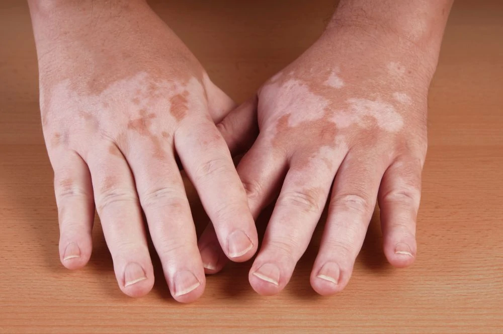
Vitiligo causes the loss of skin pigment (melanin). It creates distinct white patches that can appear anywhere, though they often start around the eyes, mouth, neck, or hands.
It can affect people of all races but is more visible on darker skin tones.
The patches are sensitive to the sun. If your child has vitiligo, you must use a strong sunscreen on the spots to prevent burns.
Experts are still studying the cause, but it is generally considered an autoimmune disorder where the body attacks its own pigment cells (2).
Tinea Versicolor
Tinea versicolor is a fungal infection. It is caused by an overgrowth of yeast that naturally lives on the skin.
What to look for:
- Patches that are lighter (or sometimes darker) than the surrounding skin.
- Mild itching.
- Fine scaling if you look closely.
The fungus prevents the skin from tanning, so these spots become very obvious in the summer. It is easily treated with antifungal creams prescribed by your pediatrician, though the color might take months to even out (3).
Yellow Discoloration (Jaundice)

Seeing your baby turn yellow is alarming, but it is one of the most common newborn conditions.
Jaundice typically appears two to three days after birth. It starts in the face and can move down the chest and tummy. It can also make the whites of the eyes look yellow.
It happens because of a buildup of bilirubin in the blood. Newborn livers are still learning how to process this waste product from red blood cells.
Take Note
Most cases are mild and resolve on their own within a week or two. Frequent feedings help your baby poop, which clears the bilirubin from their system.
When to call the doctor:
- If jaundice appears within the first 24 hours of life.
- If the baby is listless, hard to wake, or not feeding well.
- If the yellow color deepens or spreads to the legs.
Severe cases need medical help to prevent brain damage. Treatments include:
- Phototherapy: Your baby lies under special blue lights (bili-lights) that break down the bilirubin.
- Exchange transfusion: In very rare, severe cases, a blood transfusion replaces the baby’s blood with fresh blood (4).
Common Birthmarks
Birthmarks are skin discolorations present at birth or appearing shortly after. They come in two varieties: vascular (blood vessels) and pigmented (cells).
1. Salmon Patch (Stork Bites & Angel Kisses)
These are pink, flat, irregular patches. They are incredibly common, affecting nearly half of all newborns.
When they appear on the forehead or eyelids, we call them “angel kisses.” These usually fade by age two. When they are on the back of the neck, we call them “stork bites.” Stork bites often persist into adulthood but are covered by hair.
They may glow brighter when your baby cries or strains. They are harmless and require no treatment.
2. Infantile Hemangioma (Strawberry Mark)
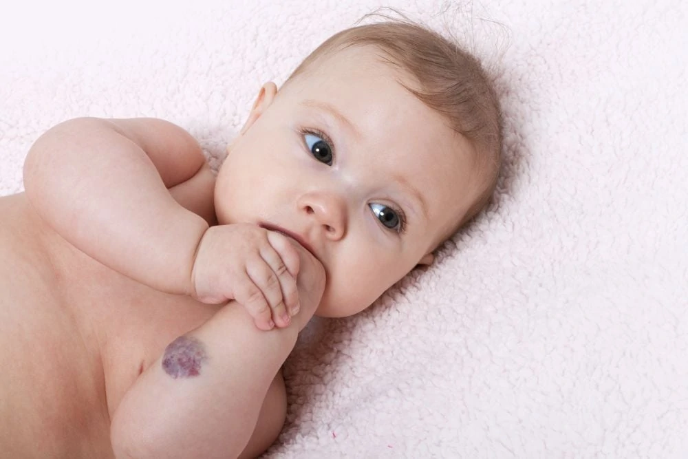
Often appearing a few weeks after birth, these look like bright red, raised bumps, much like a strawberry.
Hemangiomas tend to grow rapidly for the first six months, then shrink (involute) over several years. Most are gone by the time a child starts school.
While usually harmless, location matters. If a hemangioma is near the eye, nose, or mouth, it can interfere with vision or breathing. In these cases, doctors might prescribe medication like propranolol to shrink it safely (5).
3. Congenital Dermal Melanocytosis (Mongolian Spots)
Formerly called Mongolian spots, these are flat, blue-gray patches that look a lot like bruises.
They are very common in babies with darker skin tones (Asian, African, Hispanic, and Mediterranean heritage). You usually find them on the lower back or buttocks.
They are completely benign and typically fade by school age (6). Because they look like bruises, it is helpful to point them out to your pediatrician at the first visit so they are documented in your baby’s medical record.
4. Port-Wine Stain (Capillary Malformation)
A port-wine stain is a flat, reddish-purple birthmark caused by swollen blood capillaries. Unlike salmon patches, these do not fade. They grow as the child grows and can darken or thicken over time.
They can appear anywhere but are common on the face.
While mostly cosmetic, port-wine stains on the forehead or eyelid are sometimes associated with Sturge-Weber syndrome, a neurological condition. Laser treatments are very effective at lightening these marks, especially when started early (7).
Baby Skin Discoloration FAQs

What causes uneven skin tone in babies?
Uneven skin tone is usually caused by immature blood circulation (acrocyanosis), thin skin showing blood vessels (mottling), or harmless birthmarks. It is normal for a baby’s skin to change color with temperature or crying as they adapt to the outside world.

When do babies get their final skin color?
Babies typically develop their permanent skin tone within the first six to 12 months. Melanin production ramps up after birth, so you may notice their complexion darkening gradually during this time.

Does breast milk affect baby complexion?
No, breast milk does not directly lighten or darken skin. However, breast milk helps clear jaundice faster by encouraging bowel movements, which removes bilirubin from the body.

Can babies have hyperpigmentation?
Yes, babies can have hyperpigmentation. This includes birthmarks like Cafe au lait spots or darker patches following a rash or scratch. These are generally harmless and often fade or blend in over time.

When should I be concerned about my baby’s skin discoloration?
Seek medical advice if discoloration is accompanied by fever, feeding difficulties, or lethargy. Also, consult a doctor if a birthmark bleeds, changes shape rapidly, or if you count more than six large brown spots.

Is it normal for a baby’s skin to look marbled?
Yes, this is called cutis marmorata. It looks like a lacy, blue or red pattern on the skin and happens when a newborn is cold. It typically disappears once the baby is warmed up.